Abdominal ultrasound
Learning chapters: Liver ultrasound
Learning liver ultrasound means finding your way around the right upper abdomen and identifying vessels by following their course. Ask more than “What is this dark structure?”: where does it come from, where does it lead, and how does its appearance change when the transducer tilts or rotates?
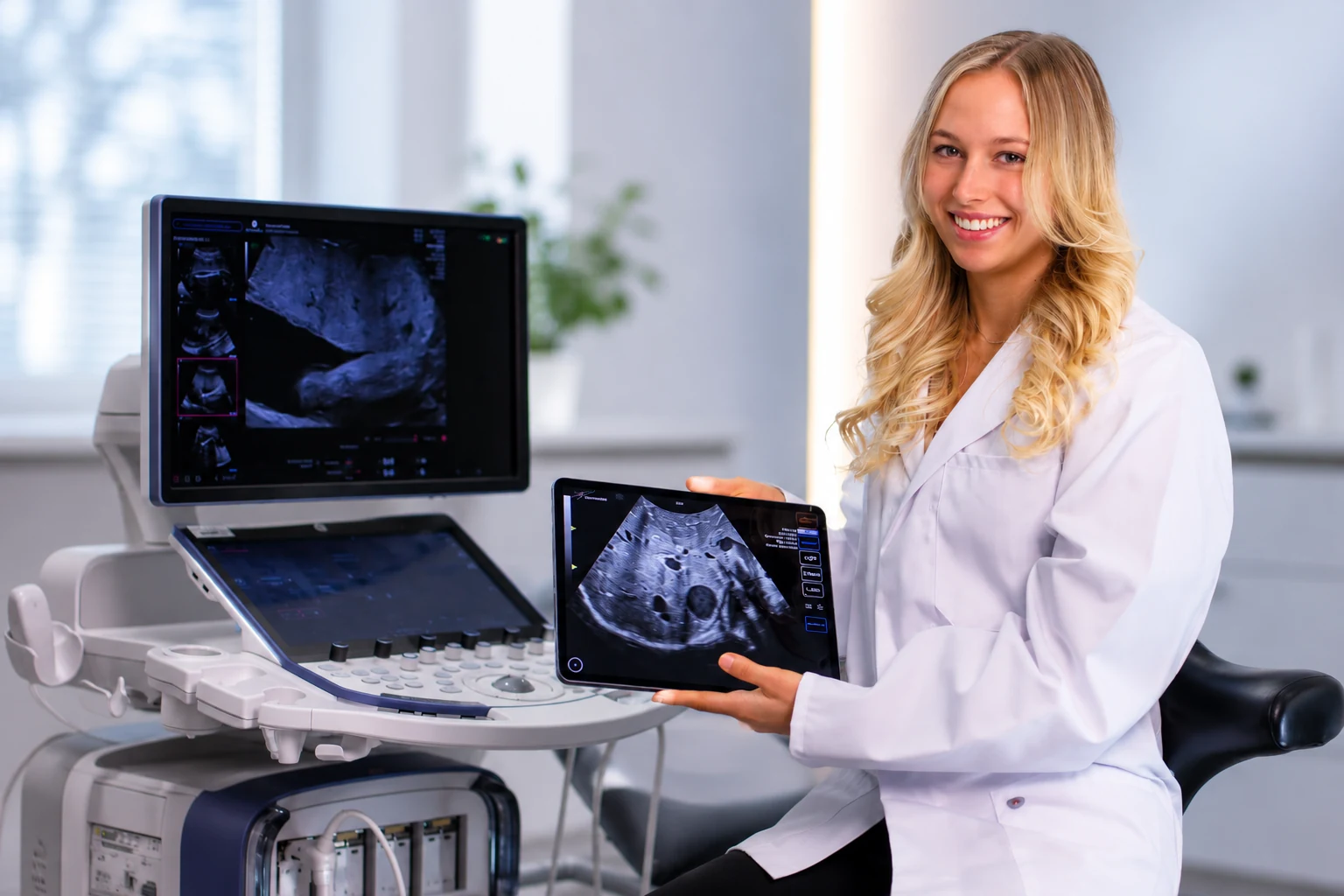
In this tutorial, Jasmin explores a physiological liver case. Scanbooster’s live labels distinguish arterial vessels, the portal vein, hepatic veins and the biliary system. The chapters below turn that examination into a guided sonography lesson with anatomical explanations and practical exercises.
- Portal vein
- Hepatic artery
- Hepatic veins
- Common bile duct
0:43Find the liver in transverse and longitudinal planes
Find the liver in transverse and longitudinal planes
Start by orienting yourself in the upper abdomen. The tutorial opens the physiological liver case under Internal Medicine → Liver. The position pictogram shows how the current scan plane relates to the body. Transverse and longitudinal sections offer different views of the same organ volume.
Use small fanning, rocking and rotating movements to explore the liver in three dimensions. Follow a structure across adjacent sections rather than naming it from a single still image. Begin with the labels switched on, then find the same structures again without the colour overlay.
Display the same vessel in two scan planes and compare its appearance with the orientation pictogram.
Check your understanding
Why should you follow the same vessel through several scan planes?
Show the explanation
A single section shows only part of the anatomy. Following a vessel through adjacent planes reveals its course and connections, helping you identify it. The orientation pictogram shows how each plane relates to the body.
1:30Locate the liver beneath the diaphragm
Locate the liver beneath the diaphragm
The liver lies mainly in the right upper abdomen beneath the diaphragm, extending partly into the left upper abdomen. The labelled diaphragm provides a landmark for the upper boundary of the liver and its relationship to the chest.
The liver has important metabolic, synthetic, storage and detoxification functions. For ultrasound orientation, its relationships to vessels and bile ducts are particularly useful. These structures provide landmarks that you can return to as you move through the organ.
Identify the diaphragm as a boundary of the liver before focusing on individual vessels.
Check your understanding
Which structure helps you identify the upper boundary of the liver?
Show the explanation
The diaphragm lies above the liver and separates the abdomen from the chest. In the tutorial, it provides an orientation landmark before individual vessels are examined.
2:16Trace the aorta, coeliac trunk and hepatic artery
Trace the aorta, coeliac trunk and hepatic artery
The tutorial starts the arterial search at the aorta. From there, it identifies the coeliac trunk and the more caudal superior mesenteric artery. Following the common hepatic artery leads towards the proper hepatic artery, which carries oxygenated blood to the liver.
The video also shows the splenic artery and the origin of the gastroduodenal artery. The proper hepatic artery divides into right and left branches. Use these branching relationships as a route map: a smaller vessel becomes easier to identify when you can trace it back to a larger trunk.
Follow the sequence aorta → coeliac trunk → common hepatic artery → proper hepatic artery.
Check your understanding
Which vessels form the arterial route from the aorta towards the liver?
Show the explanation
Follow the aorta to the coeliac trunk, then the common hepatic artery and proper hepatic artery. The proper hepatic artery divides into right and left branches supplying the liver.
4:00Recognise the portal vein and its bifurcation
Recognise the portal vein and its bifurcation
The portal vein carries nutrient-rich blood from the unpaired abdominal organs to the liver. It is labelled light blue in the tutorial. At the porta hepatis, it branches towards the right and left liver lobes. Rotating the transducer helps bring this branching pattern into view.
The tutorial compares the bifurcation to a leaping deer. The important skill is to follow the vessel continuously, rather than rely on a memorable shape alone. Distinguish portal venous inflow from hepatic venous outflow: both systems are within the liver, but they have different anatomical connections.
Follow the main portal vein to its branches, then locate the separate route from the hepatic veins to the vena cava.
Check your understanding
Does the portal vein carry blood towards the liver or away from it?
Show the explanation
It carries nutrient-rich blood from the unpaired abdominal organs towards the liver, branching at the porta hepatis towards the right and left lobes. The hepatic veins provide a separate route for blood leaving the liver.
4:51Find the hepatic veins and inferior vena cava
Find the hepatic veins and inferior vena cava
Blood leaves the liver through the hepatic veins. The right, middle and left hepatic veins drain into the inferior vena cava. Their converging arrangement forms the hepatic venous star shown in the tutorial.
The video uses an oblique upper abdominal plane with approximately 45 degrees of probe rotation. The anatomical connection matters more than a fixed angle. Locate the venous confluence and adjust the plane until you can follow the vessels as they enter the inferior vena cava.
Identify the right, middle and left hepatic veins, then show the larger vessel into which they drain.
Check your understanding
Which vessels form the hepatic venous confluence, and where do they drain?
Show the explanation
The right, middle and left hepatic veins converge towards the inferior vena cava. This is the hepatic venous star shown in the tutorial. The portal vein bifurcation belongs to a different vascular system.
5:39Identify the gallbladder and common bile duct
Identify the gallbladder and common bile duct
The gallbladder lies beneath the liver and stores and concentrates bile. Its cystic duct joins the common hepatic duct to form the common bile duct, or ductus choledochus. This extrahepatic duct carries bile towards the duodenum.
A bile duct is not immediately visible in every plane. The tutorial starts with the gallbladder, then uses changes in probe angle and rotation to explore the surrounding anatomy. An oblique shoulder-to-umbilicus plane is also discussed. Do not identify a dark tubular structure as a duct solely from its appearance; check its anatomical course.
Find the gallbladder first, then follow the neighbouring duct and vessel structures through adjacent planes.
Check your understanding
Which two ducts join to form the common bile duct?
Show the explanation
The common hepatic duct joins the cystic duct to form the common bile duct, or ductus choledochus. It carries bile towards the duodenum. The gallbladder connects through the cystic duct.